[Abstract and figures of
K. Isari, H. Yoshida, T. Gejo, E. Kobayashi, K. Mase, S. Nagaoka, K. Tanaka
J. Vac. Soc. Jpn. 46 (2003) 377-384 (in Japanese).
Uploaded with the permission of Vacuum Society of Japan.]
Construction and Evaluation of Coaxially Symmetric Mirror Electron Energy Analyzer with High Sensitivity, and Its Application to Coincidence Spectroscopy
Kouji ISARI*1, Hiroaki YOSHIDA*1, Tatsuo GEJO*2, Eiichi KOBAYASHI*3, Kazuhiko MASE*3, Shin-ichi NAGAOKA*4, Kenichiro TANAKA*1
*1Graduate School of Science, Hiroshima University,
1-3-1 Kagamiyama, Higashi-Hiroshima 739-8526, Japan
*2Institute for Molecular Science, 38 Nishigounaka, Myodaiji-cho, Okazaki 444-8585, Japan
*3Institute of Materials Structure Science, 1-1 Oho, Tsukuba 305-0801, Japan
*4Chemistry Group, Department of Fundamental Material Science, Faculty of Science,
Ehime University, 2-5 Bunkyo-cyo, Matsuyama 790-8577, Japan
We have developed a coaxially symmetric mirror electron energy analyzer with a high sensitivity, which was originally proposed by Kai Siegbahn in 1997. The analyzer, however, had a weak point; i.e. the performance is degraded by disturbance of the electric field near the end plates. We have improved the point by adopting compensation electrodes. Simulation with SIMION 3D version 7.0 predicts that the energy resolution (E/ΔE, FWHM) would be 300 for a pointed electron source and a solid angle of 1.2 sr. This value is about 3 times as much as the predicted value for the cylindrical mirror analyzer (CMA) used in previous EICO apparatuses. The resolution of the electron energy analyzer was obtained at UVSOR BL-2B1 by measuring an Au:4f photoelectron spectrum of a gold film. The actual energy resolution was evaluated to be 110-130. Some improvements and applications to coincidence spectroscopy are also described.

Fig. 1 Auger stimulated ion desorption mechanism. When a surface is irradiated by X-rays (or synchrotron radiation) ion desorption is induced by the following three-step processes, i.e., 1) formation of a core-hole by a core-electron emission (〜0.1 fs) , 2) formation of a two-hole state by an Auger transition (1〜10 fs), 3) ion desorption induced by Coulomb repulsion of the two holes and by electron missing from the bonding orbitals (10〜100 fs).
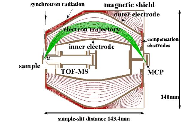
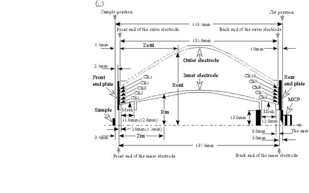
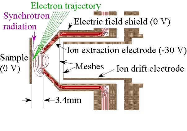
Fig. 4 Cross section of the entrance of the TOF-MS in the coaxially symmetric mirror electron energy analyzer. The isoelectric lines with 3 V step and trajectories of electrons with a kinetic energy of 181.6 eV for emission polar angles of 49°〜63° with 2° step are also shown based on the simulation with the SIMION 3D version 7.0.
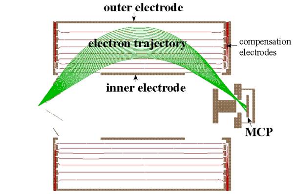
Fig. 5 Cross section of the cylindrical mirror electron energy analyzer (CMA) used for a previous electron-ion coincidence analyzer. The CMA consists of an inner electrode, an outer electrode, compensation electrodes, and MCP. The isoelectric lines simulated by the SIMION 3D version 7.0 are shown for the inner electrode at 0 V and outer electrode at -100 V. Electron trajectories are shown for the electron kinetic energy of 145.0 eV and the emission polar angles of 30°〜54° (the solid angle is 1.1 sr).
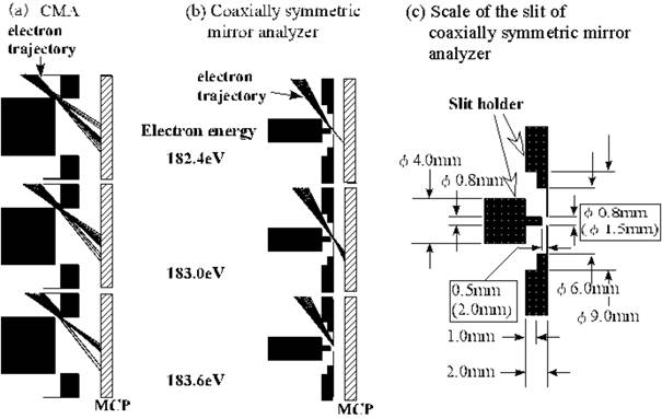
Fig. 6 Comparison of the electron trajectories around the slit of (a) the CMA and (b) the coaxially symmetric mirror analyzer. The electron kinetic energies are 182.4, 183.0 and 183.6 eV. The diameter of the slit and the width of the space in front of the slit are φ10.0 mm and 1.0 mm for the CMA, and φ0.8 mm and 0.5 mm for the coaxially symmetric mirror analyzer. (c) Scale around the slit of the coaxially symmetric mirror analyzer. The size in parentheses is for the analyzer with a large solid angle (see Sec. 6).
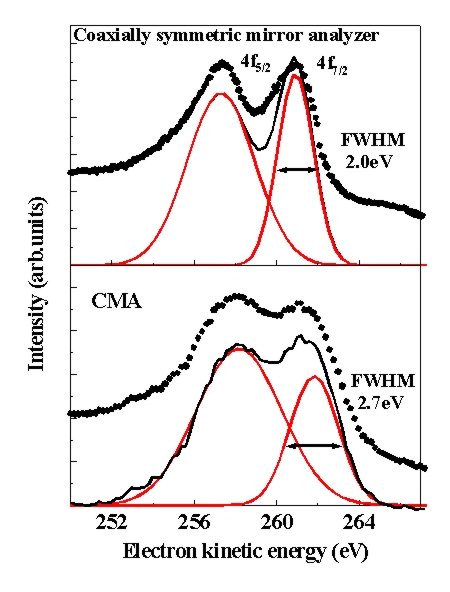
Fig. 7 Photoelectron spectra of a gold film in the Au:4f region. Based on Gaussian curve fitting the energy resolution was estimated to be E/ΔE = 130 at the 4f7/2 peak, the full width at half maximum (FWHM) of which was 2.0 eV.
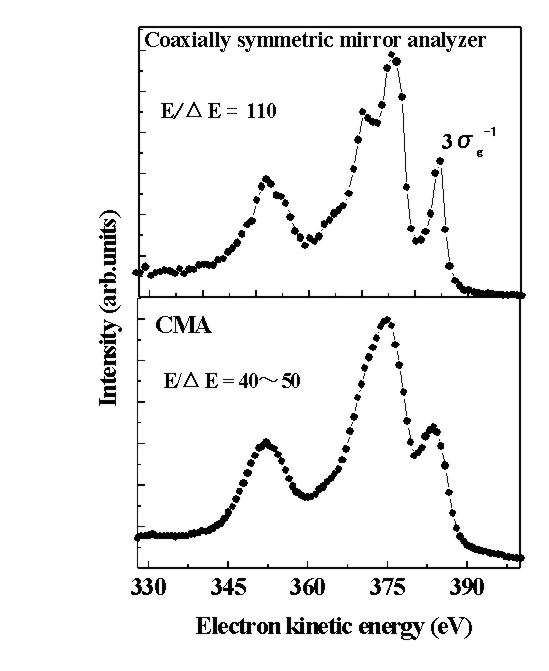
Fig. 8 Resonant Auger spectra of N2 gas at the π*←1s resonance. The energy resolution was estimated to be E/ΔE = 110 from the FWHM of the 3σg-1 peak.