[Abstract and figures of
E. Kobayashi, K. Isari, M. Mori, K. Mase, K. Tanaka, K. Okudaira, and N. Ueno,
J. Vac. Soc. Jpn. 47 (2004) 14-21 (in Japanese).
Uploaded with the permission of Vacuum Society of Japan.]
Construction and Evaluation of Polar-Angle-Resolved Miniature Time-of-Flight Ion Mass Spectrometer, and Its Application for Electron-Ion Coincidence Spectroscopy
Eiichi KOBAYASHI*1,*2, Kouji ISARI*3,*4, Masanobu MORI*5, Kazuhiko MASE*1, Koji OKUDAIRA*5,*6, Kenichiro TANAKA*3, Nobuo UENO*5,*6
*1(Institute of Materials Structure Science, 1-1 Oho, Tsukuba 305-0801, Japan)
*3(Graduate School of Science, Hiroshima University, 1-3-1 Kagamiyama, Higashi-Hiroshima 739-8526, Japan)
*5(Graduate School of Science and Technology, Chiba University, 1-33 Yayoi-chyo, Inage-ku, Chiba 263-8522, Japan)
*6(Faculty of Engineering, Chiba University, 1-33 Yayoi-chyo, Inage-ku, Chiba 263-8522, Japan)
Coincidence measurement of energy-selected electrons and mass-selected ions (electron-ion coincidence (EICO) spectroscopy) is a powerful technique to clarify ion desorption mechanism induced by electron transitions, because excited states leading to ion desorption is directly identified. Information on the coincidence ions, however, is limited to the mass and the yield so far. In order to obtain information on kinetic energy and desorption polar angle of ions, we have developed a new polar-angle-resolved miniature time-of-flight ion mass spectrometer (TOF-MS), which can be used for EICO spectroscopy. The TOF-MS consists of a shield for electric field, an ion drift electrode with three meshes, and microchannel plates with three concentric anodes. By using SIMION 3D version 7.0 we simulated ion trajectories of H+ for the TOF-MS with a drift bias of –30 V. The results show that the desorption angles of H+ with a kinetic energy of 3 eV detected by the innermost anode, the intermediate anode, and the outermost anode are 0°〜17°, 22°〜48°, and 57°〜90°, respectively. The performance of the TOF-MS was evaluated by measuring H+ yield spectra and Auger electron - H+ coincidence spectra of condensed water (H2O).
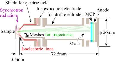
Fig. 1 Cross section of a miniature TOF-MS with a single anode. The TOF-MS consists of a shield for electric field, an ion extraction electrode with a mesh, an ion drift electrode with two meshes, and microchannel plates (MCP). The isoelectric lines with 3 V step between the sample and the ion extraction electrode, and trajectories of ions from a pointed source with a kinetic energy of 2 eV for desorption polar angles of 0°〜45° with 5° step are also shown based on the simulation with the SIMION 3D ver. 7.0. The voltage of the sample is -0 V, that of the ion extraction electrode is –30 V, that of the ion drift electrode is -300 V and that of the MCP entrance is -2000 V.
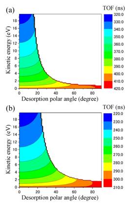
Fig. 2 TOF contour map of H+ for the miniature TOF-MS with a single anode as a function of the desorption polar angle and the kinetic energy. The geometry is the same to that of Fig. 1. The voltage of the sample is 0 V, that of the ion extraction electrode is -30 V, those of the drift electrode are (a) -300 V and (b) -1000 V, and that of the MCP entrance is -2000 V.
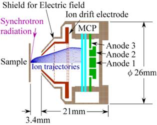
Fig. 3 Cross section of a polar-angle-resolved miniature TOF-MS with three anodes. The TOF-MS consists of a shield for electric field, an ion drift electrode with three meshes, MCP, and three concentric anodes. Trajectories for ions with a kinetic energy of 2 eV from a pointed source are shown for the desorption polar angles of 0°〜90° with 5° step based on the simulation with SIMION 3D ver. 7.0. The voltage of the sample is 0 V, that of the drift electrode is -30 V and that of the MCP entrance is -2000 V.
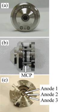
Fig. 4 Photographs of the polar-angle-resolved miniature TOF-MS with three concentric anodes. (a) the front view, (b) the side view, and (c) the three concentric anodes.
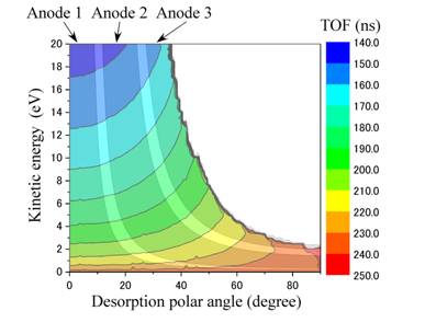
Fig. 5 TOF contour map of H+ for the polar-angle-resolved miniature TOF-MS with three anodes as a function of the desorption polar angle and the kinetic energy. The geometry and the electrode voltages are the same to those of Fig. 3.
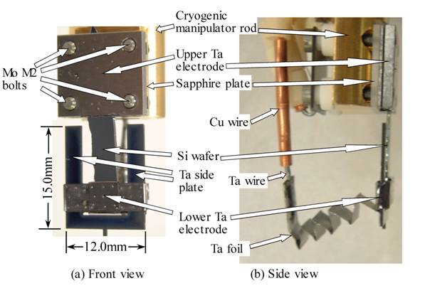
Fig. 6 Photographs of a silicon wafer holder with Ta electrodes for direct current heating.
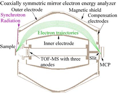
Fig. 7 Cross section of an electron – polar-angle-resolved-ion coincidence analyzer which consists of a coaxially symmetric mirror electron energy analyzer and the polar-angle-resolved miniature TOF-MS with three anodes. The details of the electron energy analyzer are described in Ref. 12.

Fig. 8 Total ion yield (TIY) with the anode 1 (solid line) and the anode 2 (dashed line), Auger electron yield (AEY, electron kinetic energy (Ekin) = 490 eV)), and TIY/AEY spectra with the anode 1 (solid line) and the anode 2 (dashed line) for condensed H2O.

Fig. 9 Auger electron spectra (solid line) and Auger electron - H+ photoion coincidence spectra (closed squares and circles) at 4a1 ← O 1s resonance of condensed H2O. The errors are estimated according to Ref. 23. The accumulation time for each coincidence signal is 640 s.

Fig. 10 Auger electron - H+ photoion coincidence spectra normalized by the total ion count for each anode at 4a1 ← O 1s resonance of condensed H2O. The errors are estimated according to Ref. 23.

Fig. 11 Auger electron - photoion time-of-flight (TOF) difference spectra at 4a1 ← O 1s resonance of condensed H2O. The accumulation time for each spectrum is 640 s.