[Abstract and figures of
K. Mase, E. Kobayashi, M. Mori, Y. Kobayashi, S. Terashima, K. Okudaira, N. Ueno,
J. Vac. Soc. Jpn. 47 (2004) 334-338 (in Japanese).
Uploaded with the permission of Vacuum Society of Japan.]
Construction and Evaluation of Miniature Cylindrical Mirror Electron Energy Analyzer (CMA), and Its Application for Auger-Photoelectron Coincidence Spectroscopy
Kazuhiko MASE*1, Eiichi KOBAYASHI*1, *2, Masanobu MORI*3, Yoshiharu KOBAYASHI*4, Shin-ichi TERASHIMA*4, Koji OKUDAIRA*3, *5, Nobuo UENO*3, *5
*1(Institute of Materials Structure Science, KEK, 1-1 Oho, Tsukuba 305-0801, Japan)
*3(Graduate School of Science and Technology, Chiba University, 1-33 Yayoi-cyo, Inage-ku, Chiba 263-8522, Japan)
*4(Mechanical Engineering Center, KEK, 1-1 Oho, Tsukuba 305-0801, Japan)
*5(Faculty of Engineering, Chiba University, 1-33 Yayoi-cyo, Inage-ku, Chiba 263-8522, Japan)
We have developed a miniature cylindrical mirror electron energy analyzer (CMA) with a diameter of 26 mm. The CMA consists of a shield for electric field, inner and outer cylinders, a pinhole, and an electron multiplier. By assembling the CMA in a coaxially symmetric mirror electron energy analyzer (coASMA) coaxially and confocally we developed an analyzer for Auger-photoelectron coincidence spectroscopy (APECS). The performance was tested by measuring Si LVV Auger – Si 2p photoelectron coincidence spectra of a Si(111) surface. Features of the APECS analyzer are as follow. 1) The coincidence signal detection efficiency of the analyzer is improved by one order of magnitude from previous ones because of the large solid angle of the coASMA and the CMA. 2) The positioning is quite easy, because the coASMA and the CMA are assembled confocally on a rod with a mechanism for xyz positioning and tilt adjustment. 3) It can be installed to a general purpose ultrahigh vacuum chamber because it is constructed on a 203-mm-outer-diameter conflat flange with a 50-mm-retractable mechanism. 4) The production cost is low because the structure is simple and the number of the parts is relatively small.
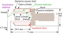
Fig. 1 Cross section of a miniature cylindrical mirror electron energy analyzer (CMA). The CMA consists of a shield for electric field, an inner cylindrical electrode with two meshes, an outer cylindrical electrode, and an electron multiplier. The incidence angle of p-polarized synchrotron radiation is 84° from the surface normal. The isoelectric lines with 10 V step and trajectories of electrons from a pointed source with a kinetic energy of 168 eV for polar angles of 29°〜41° with 2° step are shown based on the simulation with the SIMION 3D version 7.0. The voltages of the sample, the inner cylindrical electrode and the entrance of the electron multiplier are 0 V while that of the outer cylindrical electrode is -100 V.
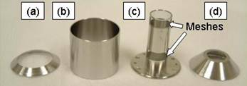
Fig. 2 Photograph of the parts of the miniature CMA. (a) The cover of the outer cylinder, (b) the outer cylinder, (c) the inner cylinder, and (d) the shield for electric field.
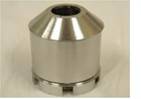
Fig. 3 Photograph of the constructed miniature CMA without an electron multiplier.
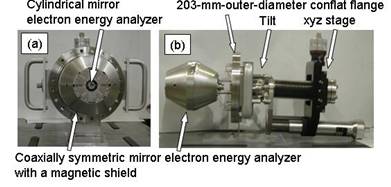
Fig. 4 Photograph of the Auger-photoelectron coincidence analyzer. (a) The front view and (b) the side view.
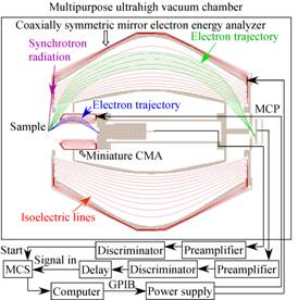
Fig. 5 Cross section of the Auger-photoelectron coincidence analyzer, which consists of the miniature CMA (Fig. 1) and a coaxially symmetric electron energy analyzer (coASMA)15). The isoelectric lines with 10 V step, trajectories of electrons with a kinetic energy of 168 eV for polar angles of 29°〜41° and trajectories of electrons with a kinetic energy of 182 eV for polar angles of 51.7°〜 67.5°are also shown based on a simulation with the SIMION 3D version 7.0. The voltages of the sample, the inner electrodes of the CMA and the coASMA, and the entrance of the electron multiplier are 0 V while those of the outer electrodes of the CMA and the coASMA are -100 V. A schematic diagram of the measurement system is also shown. The abbreviations used are as follows: MCP, microchannel plates; MCS, multichannel scaler.
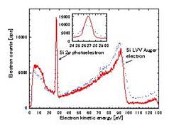
Fig. 6 Photoelectron and Auger electron spectra of a clean Si(111) surface simultaneously measured by the coaxially symmetric electron energy analyzer (solid line) and the miniature CMA (dotted line) at photon energy (hν) of 130 eV. The inlet shows the expanded Si 2p photoelectron spectra.
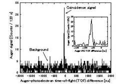
Fig. 7 Time-of-flight (TOF) difference spectrum of Si LVV Auger started by Si 2p photoelectron for a Si(111) surface measured with the Auger-photoelectron coincidence analyzer at hν = 130 eV. The Si 2p photoelectrons with a kinetic energy of 26.9 eV are detected by the coaxially symmetric mirror electron energy analyzer, while Si LVV Auger electrons with a kinetic energy of 91 eV are detected by the miniature CMA. The TOF of the Si 2p photoelectron is 65-79 ns while that of Si LVV Auger is 7-9 ns. The photoelectron and Auger counts are 2094 ± 33 and 1677 ± 41 cps, respectively. The accumulation time is 120 s. The Auger-photoelectron coincidence count is 103 ± 10 (0.86 ± 0.08 cps). The inlet shows the expanded coincidence signal.
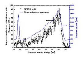
Fig. 8 Squares are Auger-photoelectron coincidence spectrum of Si(111) measured by the Auger-photoelectron coincidence analyzer at hν = 130 eV. The solid line is the singles electron spectrum collected at the same time. The accumulation time is 120 s.