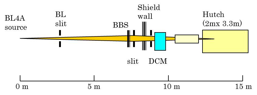
Schematic view of the beamline
BL-4A Trace element analysis/X-ray microbeam station
Contact person:Yasuhiro Niwa
5444(PHS:4942)
yasuhiro.niwa@kek.jp
Yoshio Takahashi (Univ. Tokyo)
1.Outline
This station is mainly used for X-ray fluorescence analysis with X-ray (semi-) microbeam including ultra trace element analysis, chemical state analysis, and elemental and chemical state mapping. Since synchrotron X-ray fluorescence analysis is highly sensitive to trace elements and has non-destructive nature, it is suitable for the analysis of the biological, biomedical, environmental and geological samples. It is beneficial for materials science as well.
A constant exit double crystal monochromator is equipped for the experiments with monochromatic X-rays. The experimental hutch is located 13 m from the source point.
Schematic view of the beamline
2. Area of Research
X-ray fluorescence analysis with X-ray (semi-)microbeam
trace element analysis / chemical state analysis (XANES) / elemental (chemical state) mapping /
3. Light Source & X-rays at sample
| Light source: Bending magnet | (σ:0.247mm(H) ×0.088 mm (V)) |
| Energy range | Monochromatic X-rays. 6-18 keV |
| Energy resolution(ΔE/E) | 10-4(monochromatic X-rays) |
| Beam size | Microbeam, 5μm × 5μm with KB optics |
| Photon flux | monochromatic beam(max) 1010 photons/s |
4. Facilities in Experimental Station
5. Main features of X-ray fluorescence analysis
1) Micorbeam analysis
2) Poly-Capillary analysis
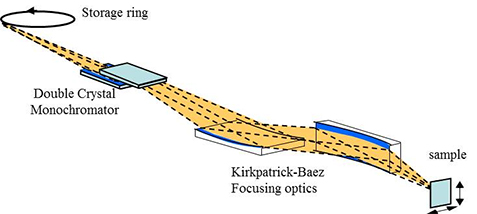
X-ray microbeam system
6. References
1) A.Iida and T.Noma: Nucl. Instrum. and Methods, B82(1993)129.
Last modified: 2014-08-25