BL-14C for X-ray imaging and multi-purpose experiments

Contact person: Kazuyuki HYODO
Office: +81-29-864-6109
kazuyuki.hyodo@kek.jp


1. Introduction

We have rearranged BL-14C at the PF experimental hall, where one can get a vertically polarized synchrotron radiation beam, to investigate X-ray phase-contrast imaging, such as X-ray phase-contrast CT using an X-ray interferometer, X-ray diffraction-enhanced imaging (DEI), DEI-CT and X-ray dark field imaging (DFI).

BL-14C1, which was dedicated to X-ray imaging, and BL-14C2, which was dedicated to X-ray diffraction experiments under high pressure and temperature, were rearranged to a large one experimental hutch, BL-14C, for X-ray imaging and other experiments using the white X-rays or the monochromatic X-rays.

A large X-ray interferometer for X-ray phase-contrast CT was installed at the back of BL-14C hutch.

2. Performance of beamline

Figure 1 shows the plan view of BL-14C at the PF experimental hall. Table 1 shows the specification for experiments. Figure 2 shows an example of experimental results.

Figure 1. Plan view of BL-14C

Table 1. Specification for experiments.
Experimental Station
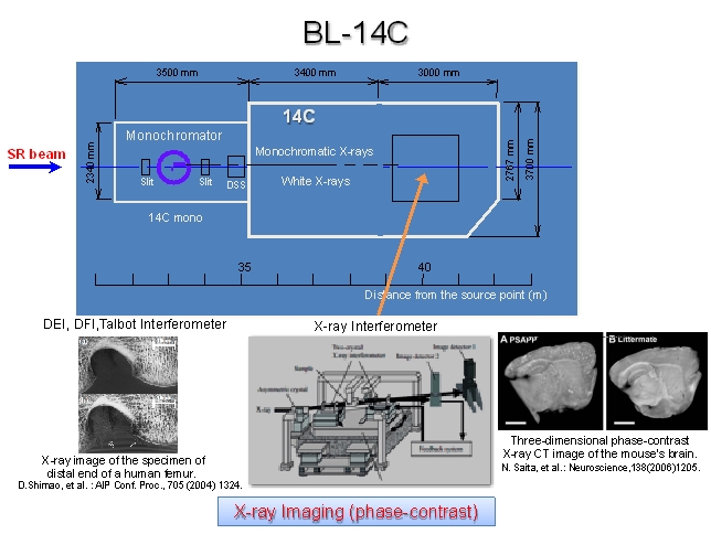
BL-14C
Source
Vertical wiggler
X-ray beam size
  • 70mm (V) by 6mm (H) at 38m
  • Typical beam size using an asymmetrical crystal:
    40mm (V) by 40mm (H) at 38m
X-ray optics
  • Si(220) double-crystal monochtomator
  • Several asymmetrical crystals are available to expand the horizontal beam size
  • X-ray interferometer
  • Laue crystals
Photon flux at 30keV
108 photons/mm2/sec
Spectral range
  • White X-rays
  • Monochromatic X-rays: 8-80keV
Experiments
  • Diffraction enhanced imaging (DEI)
  • Dark-field imaging (DFI)
  • DEI-CT
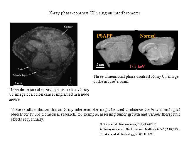
  • Imaging using an X-ray interferometer
  • Talbot interferometer

Figure 2. Results of a typical experiment at BL-14C


Last modified : 5 Apr 2013