Fig. 1
Contact person: Takumi Kikegawa
OFFICE: +81-29-846-5698
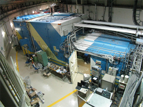
In the beginning of January 2009 new beamline AR-NE1A started its commissioning of the X-ray optical system without installation of the horizontal focusing mirror. During the commissioning period some high pressure diffraction experiments were carried out as a trial. In the end of May, the whole beamline was constructed and the user operation was taken place. First figure shows the over view of beamline AR-NE1. Main hutch(right) Experimental Hutch (Left).
Fig. 1
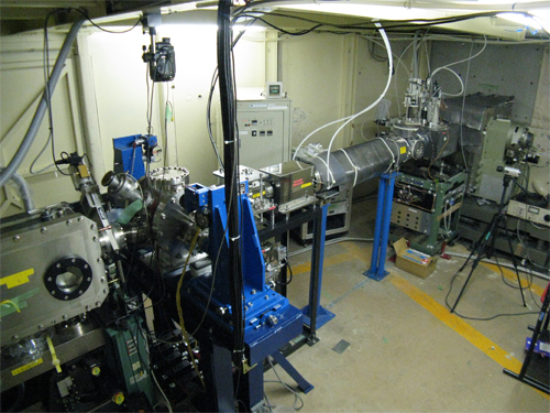
Fig. 2
The figure 2 shows optical components in the main hutch. The right side is upstream of the beamline. From right to left, a double crystal monochromator, 4D slits, high resolution monochromator, vertical focusing mirror can be seen in this picture.
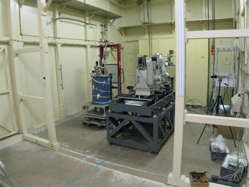
Fig. 3
Inside the experimental hutch. Experimental system of Moessbauer spectroscopy can be seen (Fig.3).
The beamline optics consists of two different kind of monochrometors and a couple of K-B type focusing mirrors. In upstream side a double crystal monochrometor was installed. It has a micro channel type water cooled 1st crystal in order to resist high heat load from the high power MPW. The other one is the high resolution monochrometor with two channel cut crystals of Si(4,2,2) and Si(12,2,2) for Mössbauer spectroscopy. The main feature of this beamline is a changeover system of focusing optics. A couple of multilayered mirrors, named tri-chromatic mirror, are newly developed for three discrete energies i.e. 14.4keV, 30keV and 50keV at a same diffraction angle. Former energy is for Nuclear resonance of 57Fe and latter two energies are for X-ray diffraction experiment with wide-Q range. Three W/C multilayer strips are fabricated on a Si substrate. Each strip is made out of 10, 40 and 60 pairs of tungsten and carbon layers with periodic length of 10.2nm, 4.2nm and 2.4nm respectively. Details and concept of the beamline AR-NE1 are shown in the previous report (*1).
Structure and Feature of AR-NE1
Schehmatic view of the beamline AR-NE1 is shown below (Fig.4). Main feature of this beamline is listed.
1.Double crystal monochromator. For high heat load from high power MPW, micro channel type water cooled crystal was adopted. Out put Energy is ranged from 7keV to 50keV, corresponding to absorption edge of Fe and high-Q for structure refinement, respectively.
2.High resolution monochrometor and graphite refraction lens. In order to get super monochromatized X-ray, i.e. dE/E~5×10-7, two channel cut crystal Si(4,2,2) and (12,2,2) are arranged for Mossbaure spectroscopy. To improve the throughput at high resolution monochromator, parallelism conversion refractive lens made of graphite will be installed in near future.
3.Trichromatic focusing Mirror. Multilayered W/C mirror is newly developed for three energy which is 14.4keV, 30keV and 50keV. Former is for Nuclear resonance of Fe and latter two are for X-ray diffraction experiment (Fig.5).
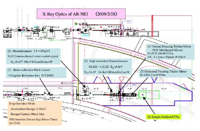
Fig. 4
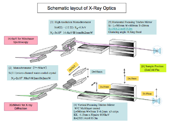
Fig. 5
Science at AR-NE1
In order to utilize the feature of high intensity and high energy single bunch SR source, X-ray diffraction or Mössbauer spectroscopy experiments under high pressure have been carried out at the first stage. Figure 6 and 7 show powder-diffraction profiles recorded on IP of same CeO2 sample in a diamond anvil cell (DAC) obtained by focused 30keV SR and higher energy 50keV SR (*2). At low temperature and high pressure condition, time-domain 57Fe nuclear resonant forward scattering (NFS) experiments has been carried out by using 14.4keV single bunch SR. Figure 8 shows quantum beat signals from 57Fe nuclei of EuFe2As2 sample in a miniature DAC (*3). As the next stage, we are planning to combine these two methods of measurement for one sample at the same pressure-temperature condition in FY2010.
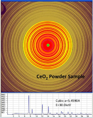
Fig.6 Diffraction profile of CeO2 sample shows many diffraction peaks. It was obtained by using normal energy (30keV) SR focused by multilayered mirrors. Incident beam was shaped by a pin hall of diameter size being 30 micron.
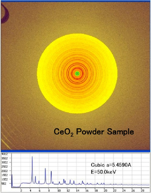
Fig.7
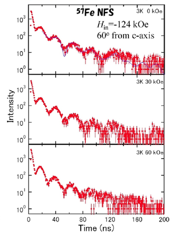
Fig. 8
(*1) Photon Factory Activity Report, 26(2009) 85
(*2) With courtesy of Prof.N.Funamori, Dr.T.Sato and Mr.D.Wakabayashi (The Unoversity of Tokyo).
(*3) NFS data by courtesy of Prof.H.Kobayashi (University of Hyogo)
Last modified:
2012-10-02
![]()